

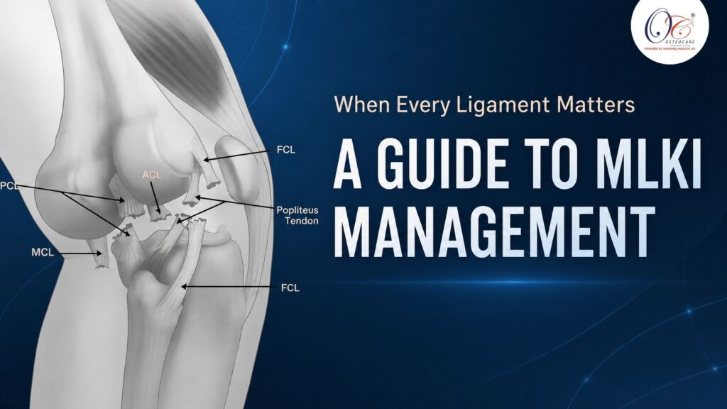
MLKI: A Comprehensive Guide to Multiligament Knee Reconstruction
Multiligament Knee Injuries (MLKIs)
are complex, often high-energy injuries involving two or more of the four major knee ligaments: the Anterior cruciate ligament (ACL), the Posterior cruciate ligament (PCL), the Medial collateral ligament (MCL), and the Lateral collateral ligament/Posterolateral corner (LCL/PLC). These injuries result in significant joint instability and represent a serious challenge in orthopedic trauma, often leading to poor functional outcomes and long-term arthritis if not managed appropriately.
Classifying Multiligament Injuries
Multiligament injuries are most commonly classified by using the Schenck classification system for knee dislocation (KD). This system categorizes the injury based on the direction of tibial displacement relative to the femur.
Schenck’s classification system for knee dislocation (KD).
| Classification | Description | Injured structures |
|---|---|---|
| KD-I | Isolated ligament injury | ACL or PCL |
| KD-II | Bicruciate ligament injury | ACL and PCL |
| KD-III-M | Bicruciate injury with medial-sided disruption | ACL, PCL, and MCL |
| KD-III-L | Bicruciate injury with lateral-sided disruption | ACL, PCL, and FCL |
| KD-IV | Combined bicruciate and collateral ligament injuries | ACL, PCL, MCL, and FCL |
| KD-V | Dislocation with periarticular fracture | ACL, PCL, MCL, FCL, and periarticular bones |
ACL: Anterior cruciate ligament, PCL: Posterior cruciate ligament, MCL: Medial collateral ligament, FCL: Fibular collateral ligament, M: Medial, L: Lateral
🔎 Assessment of MLKIs
A thorough and systematic assessment is critical due to the potential for limb-threatening vascular inury and associated nerve damage.
| Assessment Phase | Component | Key Actions & Tests | Rationale & Significance |
|---|---|---|---|
| 1. Primary Survey (Limb-Threatening) | Neurovascular Status | Immediate priority. Assess before detailed ligamentous exam. | To rule out vascular injury or compartment syndrome, which are surgical emergencies. |
| Vascular Assessment | • Palpate distal pulses (dorsalis pedis, posterior tibial).• Perform Ankle-Brachial Index (ABI).• Assess for “5 Ps” of ischemia: Pain, Pallor, Paresthesia, Pulselessness, Paralysis. | • A palpable pulse alone is not sufficient to rule out an intimal tear.• ABI < 0.9 is a red flag.• Any sign of ischemia mandates immediate vascular surgery consultation and CT Angiography (CTA). | |
| Neurological Assessment | • Peroneal Nerve: Check for foot drop (motor) and sensation on the dorsal foot.• Tibial Nerve: Check for plantar flexion (motor) and sensation on the sole. | The peroneal nerve is the most commonly injured nerve in MLKIs, especially with lateral/PLC injuries. | |
| 2. Secondary Survey (Structural) | Physical Examination | Performed once the limb is stable. Best done with patient relaxed; exam under anesthesia may be required. | To systematically identify all injured structures and classify the pattern of instability (e.g., KD-I to KD-V). |
| Ligamentous Stability Tests | • PCL: Posterior Sag Sign, Posterior Drawer Test.• ACL: Lachman Test, Anterior Drawer Test.• MCL/Medial Side: Valgus Stress Test at 0° & 30° flexion.• LCL/PLC/Posterolateral Corner: Varus Stress Test at 0° & 30° flexion, Dial Test at 30° & 90°. | • Testing at 0° vs. 30° helps differentiate isolated ligament vs. combined (capsular) injury.• The Dial Test helps distinguish PLC injury from other causes of external rotation. | |
| 3. Diagnostic Confirmation & Planning | Imaging | Essential for confirming diagnosis, classifying injury, and surgical planning. | |
| Plain Radiography | • Standard Anteroposterior (AP) and Lateral views.• Look for: Segond fracture (lateral capsular avulsion), tibial plateau fractures, fibular head avulsion. | Rules out associated fractures and confirms dislocation type (e.g., tibiofemoral dislocation). A weight-bearing view is typically avoided initially. | |
| Stress Radiography | • Varus/Valgus Stress Views. | Quantifies the degree of joint line gapping (in mm), useful for grading collateral ligament injuries (e.g., Grade III MCL) and guiding repair vs. reconstruction decisions. | |
| Magnetic Resonance Imaging (MRI) | • Gold standard for soft tissue assessment. | Precisely identifies all torn ligaments, meniscal injuries, cartilage status, and bone bruises. Crucial for pre-operative planning to determine which structures require repair or reconstruction. |
Surgical Reconstruction
The surgical approach is tailored to the specific injury pattern (KD type). The general principle is to reconstruct the cruciate ligaments (ACL/PCL) first to restore central stability, followed by the collateral ligaments/PLC.
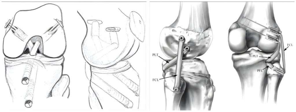
- ACL/PCL Reconstruction: These are typically performed anatomically, drilling tunnels through the femur and tibia.
- Collateral Ligaments: The MCL is often repaired or reconstructed. The PLC is generally reconstructed with two separate tendon bundles (e.g., FCL and Popliteus) to recreate its complex anatomy.
Most MLKIs are addressed in a single-stage surgery, where all damaged ligaments are reconstructed in one operation. However, in some cases with severe bony fractures or irreparable vascular injuries, a staged surgery may be necessary, addressing the most critical issues first and the ligaments later.
Avoiding Tunnel Convergence
A critical technical challenge is Avoiding Tunnel Convergence. This occurs when multiple drill tunnels (for ACL, PCL, and sometimes collateral ligaments) cross or compromise each other within the femur or tibia, which can lead to inadequate fixation or tunnel enlargement.
- Strategies to Avoid Convergence:
- Staging the surgery: Performing ACL/PCL first, then addressing the collaterals.
- Using different portals/angles: Adjusting the entry points and angles for the drill guides to maximize bone bridges between tunnels.
- Anatomic Placement: Precise, anatomic tunnel placement is the best defence against convergence.
- Femoral Fixation: Using independent suspension devices (e.g., buttons) instead of cross-pin fixation near the femoral tunnels can save space.
The Recovery Timeline: A Phased Approach
| Phase | Timeline | Key Goals | The “Do Not” List |
|---|---|---|---|
| Phase 1: Protection | Weeks 0-6 | Control pain/swelling, Protect grafts, Activate muscles. | NO WEIGHT-BEARING. Brace locked straight. |
| Phase 2: Motion | Weeks 6-12 | Restore Range of Motion, Begin gentle strengthening. | NO FORCED STRETCHING. Avoid pivoting. |
| Phase 3: Strength | Months 3-9 | Build strength & endurance, Improve balance & control. | NO HIGH-IMPACT. Listen to your physio! |
| Phase 4: Return | Months 9-18+ | Sport-specific training, Gradual return to activity. | NO RUSHING. The last thing you need is a re-injury. |
Conclusion
Multiligament knee injuries represent a complex challenge requiring a team-based approach. Advances in imaging, surgical techniques, fixation innovations, and structured rehabilitation have transformed the way surgeons treat MLKIs today. With precise diagnosis, meticulous reconstruction, and dedicated rehabilitation, patients can achieve stable, functional knees and often return to their previous level of activity.